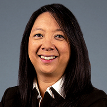
Paolo Antonio Silva, MD
-
Academic Faculty, Clinical Provider, Researcher
-
Adult Diabetes, Eye Care
-
Vascular Cell Biology
Ophthalmologist
Assistant Chief of Telemedicine, Beetham Eye Institute
Assistant Investigator
Associate Professor of Ophthalmology, Harvard Medical School
Dr. Silva's primary expertise lies in the fields of ocular telehealth for diabetic retinopathy, ultrawide field (UWF) retinal imaging and electronic medical record review. Dr. Silva has contributed 79 scientific publications with 26 original articles (17 as either first or last author), 16 study group publications, 25 review articles, 2 clinical guidelines and 9 book chapters. His work and efforts have been recognized with 20 national and international awards that include Young Clinician Award from the Center for Integration of Medicine and Innovative Technology and Eleanor and Miles Shore 50th Anniversary Fellowship Program for Scholars in Medicine, Harvard Medical School. He has been recognized by Philippine President Benigno S. Aquino III as one of The Outstanding Young Men Awardees for Medicine in 2013 and the Presidential Award for Filipinos Overseas in 2014. Dr. Silva directs telemedicine and retinal imaging research programs that have led to collaborative efforts in the Philippines with Joslin and the Diabetic Retinopathy Research Network (DRCR.net). Presently, the Philippine reading center is prospectively evaluating UWF angiograms and retrospectively evaluating optical coherence tomography scans from clinical trials conducted by DRCR.net.
As Assistant Chief of Telemedicine, Dr. Silva directs efforts of the Joslin Vision Network (JVN) in Boston, provides support to over 90 clinical sites through the Indian Health Service and develops collaborative efforts with both industry and clinical programs internationally. Outcomes data derived from the JVN have led to key publications describing novel approaches to telemedicine and better understanding of telemedicine outcomes. These contributions include elucidating the role of image evaluation at the time of telemedicine imaging and the evaluation of various automated and low light imaging systems.
Since 2012, Dr. Silva has served as Associate Retina Fellowship Program Director at Joslin. He has mentored over 24 retina fellows, researchers, visiting scholars and medical students for various countries. He serves as Faculty in the HMS Patient-Doctor II course at Harvard Medical School and an examiner at the Ateneo School of Medicine and Public Health, Philippines. He has been selected as one of the best mentors in 2016 at The Medical City and the Ateneo School of Medicine. He has conducted multiple instructional courses on ocular telehealth at national and international meetings.
His administrative emphasis has been on fostering greater clinical innovation and the development of infrastructure that will facilitate and nurture such innovation. Dr. Silva is actively involved in the Electronic Medical Record Development at Joslin and at The Medical City in the Philippines. The program at the Medical City was awarded recognition for Innovations in Healthcare IT during the Asian Hospital Management Awards 2014.
College: University of the Philippines, Manila, 1997
Medical School: University of the Philippines, Manila, 2002
Internship: Philippine General Hospital, 2001-2002
Residency: Department of Ophthalmology and Visual Sciences, Philippine General Hospital, 2003-2006
Fellowship: Joslin Diabetes Center, Harvard Medical School, 2007-2009
Dr. Silva's publications on UWF imaging have described its ability to detect posterior and peripheral diabetic retinopathy, its potential to improve efficiency of telemedicine programs and determine patients at exceptional risk for retinopathy progression. These papers have been recognized as among the top practice-changing publications in September 2012 by the American Academy of Ophthalmology and again in April 2015. His work has allowed the adoption of the UWF imaging technology with a reduction in the ungradable rate through undilated pupils by 75% with a present ungradable rate of less than 3%. This approach has reduced image evaluation time by 25%, increased the identification of retinopathy by 20% and increased the identification of sight-threatening retinopathy by 40%. Furthermore, the presence and predominance of these peripheral changes is predictive of retinopathy progression and the development of advanced sight-threatening retinopathy by nearly 4 and 5 fold respectively.

Related Experts


